


Time-lapse footage reveals cardiac cells in a mouse embryo begin to organise themselves during early development
The moment a heart begins to form has been captured in extraordinary time-lapse images for the first time.
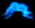
The footage reveals cardiac cells in a mouse embryo begin to spontaneously organise themselves into a heart-like shape early in development. Scientists say the technique could provide new insights into congenital heart defects, which affect nearly one in 100 babies.
More Stories
52 tiny annoying problems, solved! (Because when you can’t control the big stuff, start small)
Australians may soon be able to download iPhone apps from outside Apple App Store under federal proposal
Les Squires obituary